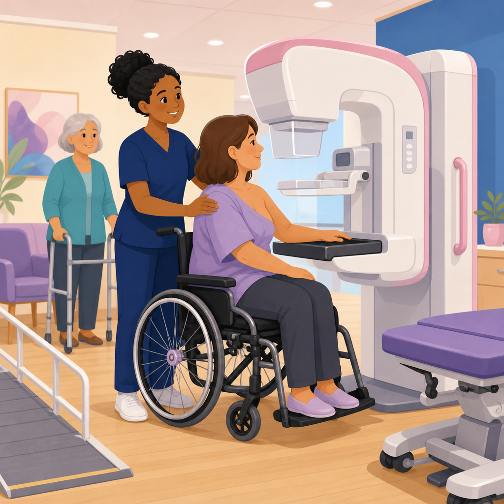
Mammography accessibility is a clinical, operational, and civil rights issue, and for patients with mobility disabilities it often determines whether breast cancer screening happens on time or is delayed until symptoms appear. In imaging departments where I have reviewed workflows, the barrier is rarely one single inaccessible machine; it is usually a chain of small failures involving parking, registration counters, changing rooms, transfer support, positioning technique, and staff assumptions. Mammography accessibility means designing screening so a person who uses a wheelchair, scooter, walker, prosthesis, or caregiver assistance can receive the same quality exam, with the same dignity, as any other patient. Mobility disabilities include limited standing tolerance, paralysis, weakness, contractures, severe arthritis, balance disorders, limb difference, chronic pain, and conditions that affect safe transfers or arm range of motion.
This topic sits squarely within medical care and accessible equipment because mammography is unusually dependent on body positioning, equipment geometry, and staff skill. Standard breast imaging requires the technologist to obtain craniocaudal and mediolateral oblique views with adequate compression, tissue inclusion, and sharpness. For many patients, that routine is straightforward. For a patient who cannot stand steadily, cannot abduct one shoulder, cannot lean forward, or cannot step close to the detector because of footrests or wheelchair frames, the standard workflow breaks down quickly. If the imaging center does not plan for these realities, the patient may be turned away, told to return with help, or receive technically limited images that trigger avoidable callbacks.
Why does this matter beyond convenience? First, delayed or incomplete screening raises the risk of later-stage cancer detection. Second, inaccessible care can violate disability rights obligations under the Americans with Disabilities Act and Section 504 in covered settings. Third, poor accommodation creates quality problems: repeat images increase discomfort, extend appointment times, and may still miss posterior tissue if positioning is compromised. A truly accessible breast imaging service is not simply a lower machine. It is a coordinated system of architectural access, adjustable equipment, transfer supports, staffing protocols, scheduling flexibility, and communication that begins before the patient arrives and continues through diagnostic follow-up if additional imaging is needed.
As a hub page for medical care and accessible equipment, this article explains the positioning barriers unique to mammography, the equipment features that improve access, the facility standards that support safe exams, and the operational practices that make accessibility reliable instead of ad hoc. It also connects mammography to the broader clinical accessibility framework: transfer equipment, height-adjustable surfaces, wheelchair clearances, patient handling methods, and competent communication. The core principle is simple: accessibility in breast imaging must preserve both equal access and diagnostic quality. If either side is missing, the service is not truly accessible.
Why Mammography Is Especially Difficult for Patients With Mobility Disabilities
Mammography is more technically demanding than many outpatient imaging exams because image quality depends on precise patient alignment against a fixed detector while the breast is drawn forward and compressed. A technologist must include posterior tissue, open the inframammary fold when possible, avoid skin folds, and position the pectoral muscle adequately on the mediolateral oblique view. Patients with mobility disabilities often face mechanical obstacles at each step. Wheelchair armrests may block access to the breast platform. Limited trunk control can make it hard to lean in. Spasticity, pain, kyphosis, or shoulder restriction can prevent the angles used in standard positioning. Even a patient who can transfer may not tolerate standing long enough for multiple exposures.
These barriers are not rare edge cases. Many breast screening patients are older adults living with arthritis, joint replacement, stroke history, Parkinsonian symptoms, neuropathy, obesity, or cardiopulmonary limits. Others are younger patients with spinal cord injuries, cerebral palsy, muscular dystrophy, multiple sclerosis, amputation, or connective tissue disorders. In practice, the accessibility question is not whether a department will see patients with mobility limitations; it is whether the department has made routine plans for them. The answer should include accessible parking and routes, doorway and turning space clearances, seating with arms, private changing space, and staff trained to ask in advance whether a patient will remain seated, transfer, or need assistance positioning the torso and arms.
There is also a persistent myth that accessibility requires sacrificing mammographic quality. In my experience, that assumption usually reflects limited training, not clinical reality. Many seated or partially supported exams can be performed successfully when the detector height lowers sufficiently, the machine head rotates appropriately, footrests or side guards are adjusted, and the technologist adapts body mechanics. The challenge is matching the exam setup to the patient’s functional abilities. Some patients can sit upright independently and only need lowered receptor height. Others require angled wheelchair placement, removable armrests, or imaging from a power chair because transfers are unsafe. A smaller subset may need diagnostic alternatives when standard views cannot be obtained despite best efforts. Planning for these scenarios is part of competent care, not an exception.
Positioning Strategies That Preserve Diagnostic Quality
Accessible positioning starts with assessment before the first exposure. The technologist should ask how the patient transfers, whether standing is possible, whether one side is weaker, how long upright tolerance lasts, and whether there is pain with shoulder elevation or torso rotation. That brief functional history often determines the safest setup more accurately than visual observation alone. For example, a patient who appears able to stand may be at high fall risk during compression release, while a patient who uses a wheelchair full time may have excellent trunk control and complete the exam seated with minimal modification.
For craniocaudal views, the primary goal remains bringing the breast onto the detector with the nipple in profile when possible and including posterior medial and lateral tissue. In a seated exam, success often depends on lowering the receptor enough that the patient can approach closely without wheelchair components blocking access. Swing-away footrests, removable armrests, and angled chair positioning can create the needed clearance. I have seen exams improve immediately when the wheelchair is turned slightly oblique rather than parked straight on, allowing the torso to come closer to the image receptor. Cushions, if medically safe to remove temporarily, may also change breast height relative to the platform.
The mediolateral oblique view is usually the hardest for mobility-disabled patients because it requires torso rotation and precise detector angle to capture posterior upper outer tissue and the pectoral muscle. Patients with limited shoulder abduction or scoliosis may need the detector angle adjusted more conservatively than standard. A technologist can often support the opposite shoulder, guide the elbow placement gently, and use short verbal cues to help the patient lean or rotate only as far as tolerated. The target is not textbook appearance at any cost; it is the best technically diagnostic image the patient can safely achieve. Compression still matters because it reduces motion and tissue overlap, but it should be explained clearly and applied in a controlled, predictable way to avoid startling a patient with pain or spasticity.
Transfers require special caution. Patients should never be manually lifted by one or two staff if a mechanical lift or transfer aid is indicated. Safe patient handling principles used elsewhere in healthcare apply in breast imaging too. If the exam can be done in the patient’s own wheelchair or scooter safely, that may be preferable to a risky transfer. If a transfer is necessary because chair dimensions prevent adequate access, staff should use established transfer protocols, enough personnel, and equipment such as transfer boards or mechanical lifts as appropriate. The imaging schedule must allow extra time. Rushing is one of the main causes of both falls and poor positioning.
| Access challenge | Common cause | Practical accommodation |
|---|---|---|
| Cannot get close to detector | Wheelchair footrests, armrests, or base block approach | Lower detector, remove or swing away rests if safe, angle chair obliquely |
| Cannot tolerate standing | Weakness, balance disorder, pain, fatigue | Perform seated exam, allow rest breaks, extend appointment slot |
| Limited shoulder motion | Arthritis, stroke, rotator cuff injury, contracture | Modify detector angle, support limb gently, accept patient-specific range |
| Unsafe transfer risk | Paralysis, obesity, spasticity, fall history | Image in personal chair when possible or use transfer equipment and protocol |
| Inadequate posterior tissue on images | Torso cannot rotate or lean into receptor | Reposition chair, lower receptor further, use incremental coaching and repeat only when needed |
Equipment Features That Improve Access
The most important mammography equipment feature for mobility access is vertical travel range. A unit that lowers sufficiently can often image patients seated in standard wheelchairs and many power chairs without compromising the technologist’s positioning options. Height adjustability sounds basic, but actual usable range varies by manufacturer and room setup. Before purchasing, facilities should test equipment with real wheelchairs, not just standing volunteers. Detector access, paddle reach, rotation controls, and clearance under the image receptor all affect whether the machine works for seated positioning in practice.
Geometry matters as much as height. Some mammography units have tube head and detector designs that allow easier lateral approach for wheelchairs; others have housings or platform shapes that create contact points too early. A center evaluating equipment should ask specific questions: Can a patient remain in a power wheelchair? Can armrests stay in place? How low does the receptor go with compression paddle attached? Does the unit permit fine angle adjustments for atypical body habitus or contractures? Are controls easy for technologists to reach while supporting a seated patient? Facilities should also examine room dimensions. A technically adjustable machine can still be inaccessible if there is inadequate turning radius, poor entry alignment, or fixed cabinetry that blocks side approach.
Accessible medical equipment principles extend beyond the mammography unit itself. Stable chairs with arms in changing rooms, grab bars where appropriate, non-slip flooring, transfer space adjacent to seating, accessible gowning areas, and scales for wheelchair users all support equitable care. If the center performs breast ultrasound or image-guided biopsy, it should also have height-adjustable exam tables and stretchers, because an accessible screening visit often leads to additional same-day or follow-up procedures. A common failure point is a center that can complete screening mammography seated but cannot accommodate the patient for diagnostic compression views, ultrasound, stereotactic biopsy preparation, or post-procedure recovery. Accessibility must cover the whole breast care pathway.
Digital breast tomosynthesis adds another layer. Many centers now use tomosynthesis routinely because it can improve cancer detection and reduce recall rates in certain populations. The accessibility question is whether the machine’s positioning and acquisition time remain manageable for the patient. Since tomosynthesis exposures may require steadiness over a brief sweep, motion control and comfort become even more important. In practice, if a patient can tolerate standard compression and maintain position for the acquisition, tomosynthesis is often feasible. If not, the facility should have protocols for adapting technique or documenting limitations without blaming the patient. Equipment should serve the patient, not the reverse.
Facility Operations, Standards, and Staff Training
Accessible mammography depends on operations as much as hardware. Scheduling scripts should ask whether the patient uses a wheelchair or scooter, whether transfer assistance is needed, and whether extra time should be booked. Front desk staff should know that asking about access needs is a service step, not an inconvenience. Online forms should provide a place to note mobility needs in plain language. Signage, reminder calls, and websites should explain available accommodations, accessible parking routes, and whether caregivers may accompany the patient into changing or imaging areas when needed.
Staff training must cover disability etiquette, safe patient handling, and mammography-specific positioning adaptations. Technologists should practice seated positioning techniques rather than improvising only when a patient arrives. In centers I have assessed, confidence rises quickly when teams build a simple protocol: review access notes before the appointment, prepare the room, identify whether a transfer is planned, gather positioning aids, and assign enough time. Quality assurance should include accessibility metrics such as incomplete exam rates for mobility-disabled patients, repeat image frequency, transfer incidents, and patient complaints about being turned away. What gets measured improves.
Standards and guidance from recognized bodies can anchor these efforts. ADA obligations shape program access and nondiscrimination. Safe patient handling guidance informs transfer decisions. Mammography quality expectations remain governed by accepted imaging practice and the Mammography Quality Standards Act environment. None of these standards excuse inaccessible care; they require centers to reconcile image quality, patient safety, and equal access through planning. The strongest programs build internal links between radiology leadership, risk management, facilities teams, and disability access coordinators so that problems are fixed systematically instead of case by case.
For organizations managing broader medical care and accessible equipment, mammography should be treated as a sentinel service. If a facility can solve access for this exam, it is usually better prepared for gynecology, dental care, imaging, infusion, and specialty outpatient services that also depend on transfer space, adjustable equipment, and coordinated staff response. Start with an access audit, identify equipment and workflow gaps, test real patient scenarios, and publish clear accommodation pathways. Better mammography accessibility improves cancer screening, patient trust, legal compliance, and overall clinical quality. Review your breast imaging service now and close the gaps before the next patient encounters them.
Frequently Asked Questions
Why is mammography accessibility for patients with mobility disabilities about more than just having an accessible machine?
Mammography accessibility is rarely determined by one piece of equipment alone. In practice, a patient’s ability to complete a screening mammogram depends on the entire care pathway working well from start to finish. That begins before the appointment with accessible parking, clear curb access, automatic doors, and a route into the imaging department that does not require navigating steep ramps, heavy doors, or long distances without seating. Once inside, registration desks, waiting areas, and changing rooms must also be designed for wheelchair users, patients who use walkers, and people who need caregiver assistance or extra time to transfer safely.
Within the exam itself, accessibility depends on whether the mammography unit can be positioned appropriately for seated or limited-standing patients, whether staff understand adaptive positioning techniques, and whether there is enough room for mobility devices, transfer assistance, and privacy. A technically “accessible” machine may still fail patients if the room is too cramped, the gowning area is inaccessible, or staff insist on a standard standing position when a modified approach would work. Delays in screening often happen because several small barriers add up: a patient cannot reach the counter comfortably, has nowhere safe to sit while changing, is rushed during positioning, or is told to reschedule because staff are unsure how to proceed. For that reason, true accessibility should be viewed as a clinical quality issue, an operational design issue, and a civil rights obligation, not simply an equipment checklist.
What positioning challenges are common during mammography for people with mobility disabilities?
The most common positioning challenges involve balance, limited ability to stand for the duration of the exam, reduced upper-body strength, difficulty leaning forward, contractures, pain, spasticity, and restricted shoulder or torso movement. Standard mammography positioning assumes a patient can stand close to the unit, rotate the body on command, grasp handles or remain stable without support, and tolerate compression while maintaining a precise posture. For many patients with mobility disabilities, those assumptions do not hold. Some may need to remain seated in a wheelchair or a specialized chair, while others can stand only briefly and require the technologist to work efficiently without sacrificing image quality.
Additional challenges can arise during specific views, especially when the exam requires body rotation, torso extension, or arm placement that is difficult after stroke, spinal cord injury, neuromuscular disease, joint replacement, amputation, or chronic pain conditions. Positioning may also be affected by involuntary movements, fatigue, or the need for caregiver assistance. The solution is not to lower imaging standards but to adapt technique thoughtfully. That can include adjusting the height of the receptor, changing the angle of approach, using seated positioning when appropriate, allowing additional time, performing one view at a time with rest breaks, and communicating each step clearly before moving the patient. Skilled technologists can often obtain diagnostic-quality images with modified methods when they understand how mobility limitations affect posture, endurance, and safe transfers.
What equipment features and room design elements make a mammography department more accessible?
Accessible mammography starts with equipment that allows meaningful flexibility. Important features include a mammography unit with a wide range of vertical travel so it can accommodate patients who are seated as well as those who are standing, controls that allow precise height adjustment, and enough open space around the unit for wheelchair positioning. The design should permit the technologist to approach the patient from different angles rather than forcing one rigid setup. Equipment should also be compatible with safe patient handling practices, particularly when a patient can pivot with assistance but cannot independently maintain position for the entire study.
Room design matters just as much. There should be adequate turning radius for wheelchairs and scooters, clear floor space free of clutter, stable seating with arm support, and changing areas large enough for a patient and assistant if needed. Doors, benches, coat hooks, mirrors, and storage should be placed at usable heights. Transfer support is another key consideration. Even when a patient does not need a full transfer, they may need grab bars, a stable chair, or space for a caregiver to remain nearby. Departments should also think beyond the exam room itself: accessible restroom access, low or adjustable check-in counters, and route planning from parking to imaging are part of the same accessibility system. When these features are absent, the patient may arrive at the machine already fatigued, uncomfortable, or discouraged, which directly affects positioning success and the likelihood of completing screening.
How can mammography staff improve the experience and image quality for patients with mobility disabilities?
Staff preparation is one of the most important factors in accessible mammography. Patients benefit when schedulers ask practical questions in advance, such as whether the patient uses a wheelchair, needs assistance with transfers, cannot stand for long, or requires extra appointment time. This allows the department to assign an appropriate room, avoid rushed scheduling, and make sure trained personnel are available. On the day of the exam, respectful communication sets the tone. Staff should ask the patient how they usually transfer, what movements are difficult, whether they prefer to remain seated, and what accommodations have helped during prior imaging visits. That approach prevents assumptions and often saves time.
Technologists can improve both comfort and image quality by using clear step-by-step instructions, moving slowly enough to avoid sudden pain or loss of balance, and adapting technique without making the patient feel like a problem to be solved. If a patient tires easily, brief pauses between views can make the exam more successful overall. If upper-extremity limitations prevent standard positioning, the technologist may need to modify body rotation, receptor height, or hand placement while still working toward adequate tissue inclusion. Staff should also know when to seek assistance rather than abandoning the exam prematurely. Training should include disability etiquette, safe patient handling, adaptive positioning strategies, and awareness of legal accessibility obligations. In many departments, the difference between a failed and successful mammogram is not advanced technology but a team that anticipates barriers, communicates well, and has practiced accessible workflows.
What should healthcare facilities do to reduce delays in breast cancer screening for patients with mobility disabilities?
Facilities should approach the problem as a system issue rather than blaming individual patients, isolated staff members, or one inaccessible room. A useful first step is to map the entire screening journey, from appointment scheduling through arrival, registration, changing, imaging, follow-up, and departure, then identify where patients with mobility disabilities encounter friction. Common failure points include inaccessible online instructions, no way to note accommodation needs during scheduling, limited appointment times for longer exams, inadequate space for mobility devices, and lack of escalation procedures when standard positioning is not possible. Solving these gaps requires operational planning, not just goodwill.
Healthcare organizations should establish clear accessibility protocols, train staff consistently, and audit real-world performance. That means reviewing whether patients are offered appropriate accommodations, whether appointment lengths are realistic, whether the physical environment supports transfers and seated positioning, and whether image completion rates differ for patients with mobility limitations. Facilities should also invite patient feedback and use it to guide quality improvement. From a legal and ethical standpoint, timely access to breast cancer screening is not optional. When screening is delayed until symptoms appear, the consequences can be serious. Departments that invest in accessible design, adaptive technique, and responsive workflow planning not only comply with disability access expectations but also improve preventive care, patient trust, and overall clinical outcomes.